Sheep Brain Dissection Lab
Follow the directions within this lab to properly dissect and expose the items on your sheep brain. Pay special attention to the items that can be found on your SHEEP BRAIN LAB COMPANION to prepare for your lab exam.
Background Information:
The sheep brain is remarkably similar to the human brain, there are however, three major differences:
1. Proportion of the Cerebrum:
For example, the sheep brain has a proportionately smaller cerebrum. The human brain has a much larger cerebrum. When observing the sheep brain, it looks much more "streamlined" due to to the smaller cerebral hemispheres.
2. Orientation of the Spinal Cord:
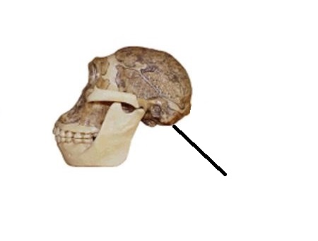
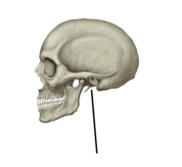
The sheep spinal cord is orientated anterior to posterior, as in any four-legged animal. The human spinal cord is orientated superior to inferior. This orientation difference has a major affect on the location of the brain stem. The sheep brain stem is located more towards the rear (posteriorly). The sheep skull, in order to compensate for this, has the foramen magnum located more towards the rear of the skull. This is because the sheep walks on all fours (quadripedalism). In humans, since we walk upright (bipedalism), the spinal cord is in a more vertical plane, thus the foramen magnum is located centrally on the bottom of the skull (inferiorly). By observing the movement of this major skull feature from the rear (as in very early human ancestors) to the current location (modern humans), scientists have been able to determine when the human species began to walk upright on two legs. Below is a picture of an early human ancestor skull and the location of the foramen magnum along side a modern human skull. Compare the differences in location of the foramen magnum. The skull on the left indicates that this early human ancestor probably did not walk totally upright yet at this time.
 |
 |
3. Size of the Olfactory Bulbs:
The sheep, being a prey animal, depends upon its sense of smell for survival in the wild. The domesticated sheep ancestors had even more pronounced olfactory bulbs than the sheep of today because to them, being able to detect a predator was vital to survival. Today's domesticated sheep have a smaller olfactory bulb due to evolution. Humans, on the other hand, we are predators and depend more on sight than our sense of smell. So the olfactory region in the human brain is much less pronounced than in other species that depend on their sense of smell for survival.
Dissection Instructions:
1. Obtain a preserved sheep brain from the bucket in the front of the classroom. Place this on your dissection tray.
2. You will need the following dissection tools to properly perform this lab:
scalpel
scissors
2 probes (1 for each lab participant)
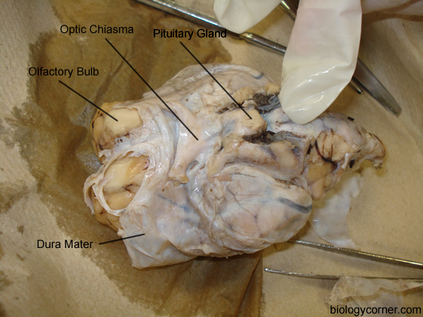
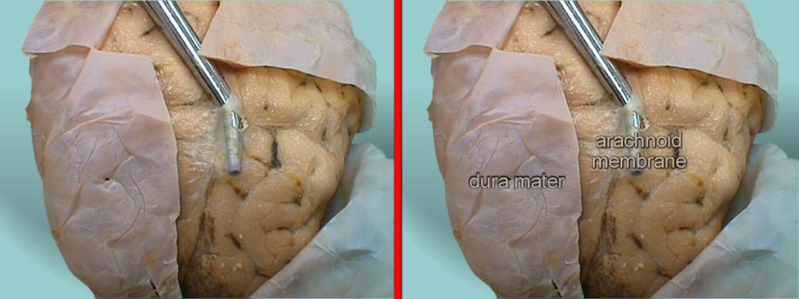
3. The sheep brain is enclosed in a tough outer covering called the dura mater. You can still see some structures on the brain before you remove the dura mater. Take special note of the pituitary gland and the optic chiasma. These two structures will likely be pulled off when you remove the dura mater. See the diagrams below to locate the following structures:
dura mater
optic chiasma
olfactory bulb
pituitary gland
arachnoid membrane

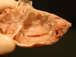
4. Using your scissors, remove the dura mater from the brain. This will not be as easy as it looks. The dura mater is a very tough membrane. As you cut into the area along the middle of the top portion of the brain, you will see how the dura mater juts downward slightly separating the right and left hemispheres of the cerebrum. This portion of the dura mater is referred to as the falx cerebri. Within the middle of the falx cerebri you will find a space or opening. This space is called the sagittal sinus. The deep opening that separates the two hemispheres is known as the longitudinal fissure. See the diagrams below to locate these structures.
Falx cerebri with the sagittal sinus opening

Longitudinal Fissue
5. After removing the dura mater from the cerebrum, carefully remove the dura mater from the cerebellum. Again, you will see a portion of the dura mater jutting down into a fissure that separates the cerebrum area from the cerebellum. The portion of the dura mater that is found here is called the tentorium cerebelli. The deep opening or fissure that separates the cerebrum from the cerebellum is called the transverse fissue. See the diagrams below to identify these structures.
Tentorium cerebelli
Transverse fissure
![]()
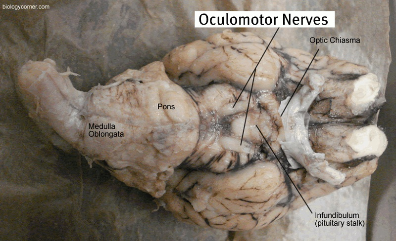
6. Once you have removed the entire dura mater, you may still be able to see the optic chiasma but the pituitary gland will probably be missing. The infundibulum (pituitary stalk) is now visible in the center. Careful dissection also reveals two other large nerves: the oculomotor nerves. Often these two nerves are removed with the dura mater, but in this image below they are still intact.
Ventral view of the brain with dura mater removed
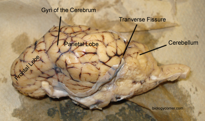
7. If you flip the brain over to the other side, you can see the cerebellum, it will be loosely attached to the cerebrum in most cases. If you did not carefully remove the dura mater you may have accidentally pulled the entire cerebellum away from the brain. The lobes of the brain are visible, as well as the transverse fissure, which separates the cerebrum from the cerebellum. The convolutions of the brain are also visible as bumps (gyri) and grooves (sulci). Use the diagram below to help you locate these items.
Dorsal View of the Sheep Brain
8. The gap between the cerebrum and the cerebellum at the transverse fissure can reveal some internal parts of the brain. In this image, a student is bending the cerebellum down to show the superior and inferior colliculi. Just behind the colliculi, the pineal gland is just barely visible.

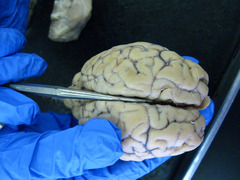
9. Using a scalpel and the longitudinal fissure as a guide, cut the brain in half so that you are left with a right side and a left side. Sharp scalpels work best for this procedure. Use the picture below as a way to see how your sheep brain should look after you cut it in half.
10. In the image below, a probe indicates the location of the lateral ventricle.

11. Once the brain is cut this way, the colliculi can also be seen from the inside and the pineal gland is revealed only if you made a very careful incision. On this image, the pineal is pinned in yellow and the pin continues on to where the colliculi have been bisected.

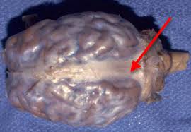
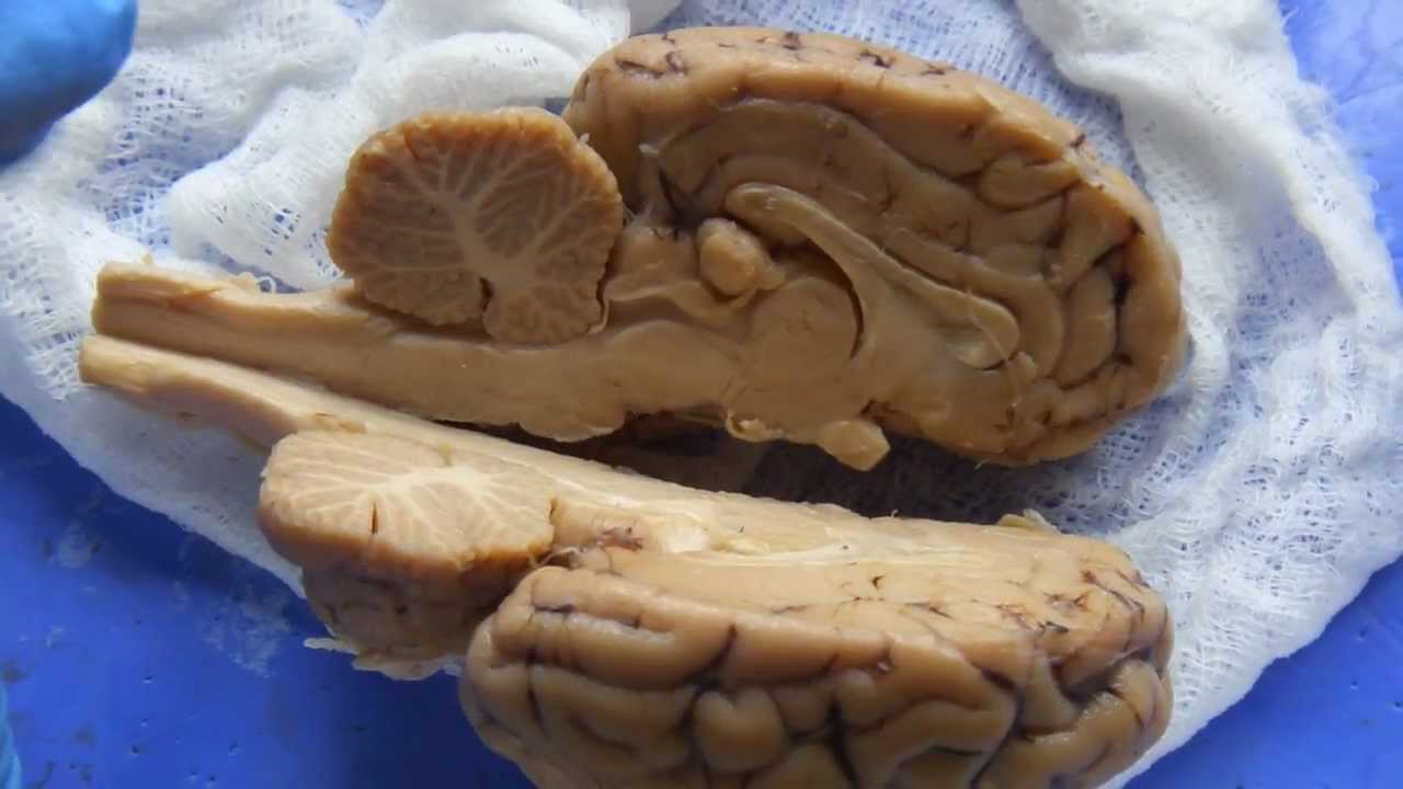
12. Other major structures are visible, here the probe indicates the arbor vitae (tree of life) found within the cerebellum. The fissure between the cerebrum and the cerebellum is called the transverse fissure. The cerebellum only loosely connects to the rest of the brain when the dura is removed.

13. The image below shows a cleanly separated brain with the major internal structures visible and labeled.

14. Finally, a section of the brain is cut to examine the difference between white matter and gray matter. Observe the picture below to see how to make this final dissection cut.

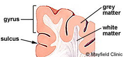
15. Once you have made the cut like in the above diagram, you should be able to see the difference between the white and gray matter, just like in the diagram below.

16. The diagram below will help you understand the difference between the gyri and the sulcus. Think of it this way: the gyri (gyrus) are the bumps and the sulci (sulcus) are the grooves between the gyri.
17. Clean up as instructed at the beginning of the class period. Complete your lab review and have it ready to hand in at the beginning of your next class period. Make sure you visit the SHEEP DISSECTION LAB COMPANION in order to prepare for the lab exam.